
The impact on patients and their health related decision making of viewing their own medical imaging results in the form of 3D accessible images
This PhD studentship is funded by the Economic and Social Research Council and University Hospitals Coventry and Warwickshire NHS Trust. The project involves exploring the impact for lay people of viewing the results of their own medical imaging (CT, MRI) in an accessible 3D format. The format of appropriate 3D images will be developed and tested by the PhD student in the Psychology Laboratory. The impact of the 3D images on the patient’s perception of their own body and their health and on health care decisions including self-management and uptake of health interventions will be evaluated qualitatively.
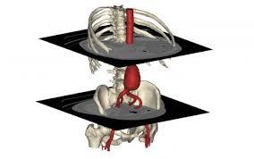
3D medical images are constructed from the digital data of a CT or MRI scan. The images are not generally used for diagnosis (radiologists use 2D grey scale), but have potential for use by clinicians and patients for increased understanding of a health issue - such as why an operation on a shoulder may or may not help symptoms and for making choices about surgical procedures. There is no additional imaging involved in their production, only manipulation of the imaging data using software that is provided by the manufacturer of the imaging machines on the powerful computers available in the imaging department. It is possible to construct the images in a huge range of formats. For example the images can be presented as if ‘peeling an onion’. For a limb this would mean presenting first the 3D image of the limb skin surface, then removing this to reveal muscle, then depending on the anatomy removing muscle to reveal further muscle and then finally presenting the bones. Another format is as if the limb has been partially dissected to reveal the pathology. The images can be presented in a range of colours, viewed from different angles or rotated. Once a format has been developed, the details of the processing can be saved and applied to subsequent patient images fairly rapidly. The construction of 3D images is not undertaken routinely in the NHS setting. Once the 3D images have been constructed, they can be saved and presented using a normal laptop.
 The research team is:
Prof Frances Griffiths (Principal Investigator)
![]()