
Warwick Medical School's collaboration with da Vinci exhibition
 Almost 500 years after Leonardo da Vinci’s death, an exhibition of the drawings in his anatomical notebooks are being displayed alongside 21st-century pictures of the same body parts - with da Vinci's sketches proving startlingly accurate.
The opening was attended by:
- Warwick Professor Peter Abrahams, representing the West Midland Surgical Training Centre (WMST) at University Hospitals Coventry and Warwickshire (UHCW);
- Dr Richard Wellings, from UHCW and a consultant radiologist,
- Mark Mobley, Warwick medical student and
- Brian Burnett of the WMSTC.
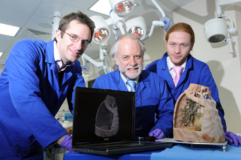
Picture captions (L-R): Mark Mobley, Professor Abrahams, Brian Burnett.
Warwick Medical School was highly commended for the work we have contributed to the exhibition.
The 3D work and CT imaging produced by WMS with joint collaboration with WMG's 3D printing lab is represented as a main feature of the exhibition.
Thousands of members of the public from across the world will visit the exhibition in the next four months and be able to appreciate these outstanding works of art.
Exhibition collaborator Peter Abrahams, Professor of Clinical Anatomy, Warwick Medical School added:
In many ways Leonardo predicted the 20th-century revolution in various medical imaging techniques. His use of cross sections and slices to show deep internal structures within the body foreshadowed the modern techniques of CT and MRI scanning. The anatomical accuracy of Leonardo's drawings has rarely ever been surpassed, and I still use them to teach surgeons and medical students today."
Leonardo da Vinci. The Mechani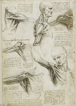 cs of Man
2 August – 10 November 2013
The Queens Gallery, Palace of Holyrood house
The Royal Collection, Edinburgh.