
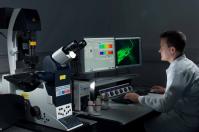
Confocal Microscopy
Conventional optical microscopes display a shallow depth of field, especially at higher magnifications. A confocal microscope uses a pinhole to collect light from the focal plane of a sample, discarding out-of-focus light to produce a clear, high resolution image.
 How does it work?
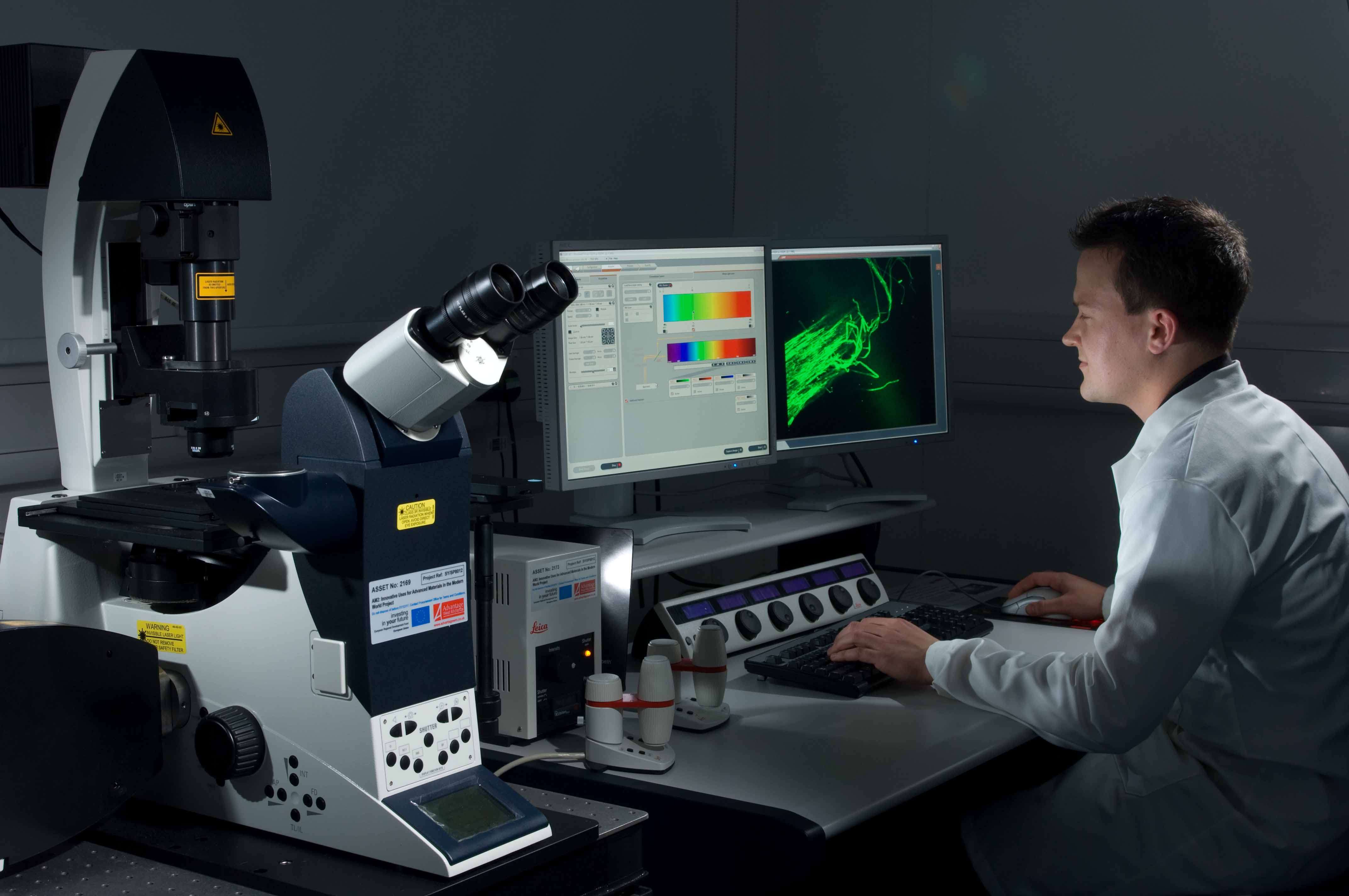
A laser is scanned across the sample, focused at a particular depth. Light from the sample passes through a small aperture (pinhole) which selects only light which contributes to an in-focus image.
Typically fluorescent species or tags are observed by selecting an appropriate wavelength of incident laser light and a suitable detection window. Multiple detectors permit several fluorophores to be monitored simultaneously.
Three-dimensional views can be obtained by stacking images from different heights in a sample, and fast scanning modes enable dynamic processes to be followed.
Applications:
Electrochemistry, neuroanatomy and neurophysiology; morphological studies of cells and tissues; resonance; energy transfer; stem cell research; photobleaching studies; lifetime imaging; multiphoton microscopy; total internal reflection; DNA hybridization; membrane and ion probes; bioluminescent proteins; epitope tagging.
Sample Handling Requirements:
Typically solid, with fluorophore(s).
Complementary Techniques:
Warwick capability:
Leica SP2 with 4 lasers, Leica SP5 with tunable white light laser.
Contact:
Claire Gerard: c dot gerard at warwick dot ac dot uk / 07385 145064
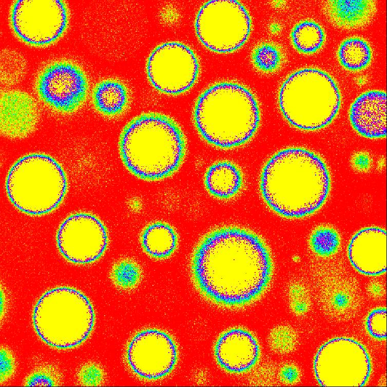
Typical results:
Status |
Availability |
|
|
Warwick collect/analyse data |
| Warwick collect data | |
| |
Available to user with expertise/ contribution |
| |
Spare capacity for collaborative research |
BOOK NOW |